In blond hair melanocytes in bulbs produce relatively few or incompletely melanized melanosomes. Normal anagen hair bulb lacks mhc i and ii hla expressed in aa allowing interaction of cytotoxic t cells with matrix cells.
The hair follicle is a dynamic organ found in mammalian skin.
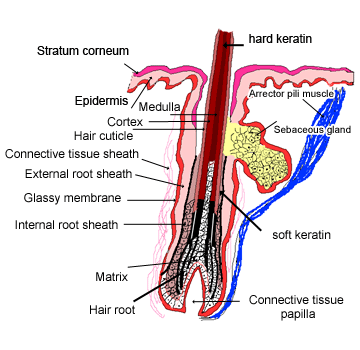
Hair bulb histology. Hair bulb hair follicle hair matrix hair papilla hair root hair shaft hypodermis internal root sheath interpapillary pegs rete pegs meissners corpuscle melanocytes of epidermis melanosomes melanin granules myoepithelial cell pacinian corpuscle papillary layer of dermis reticular layer of dermis sebaceous gland stratum basale stratum corneum. The histology of your hair can vary slightly depending your ethnicity being influenced by race and genes. It has three inner layers forming the hair shaft.
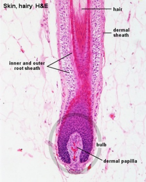
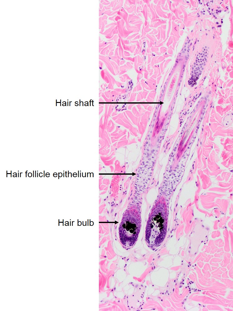
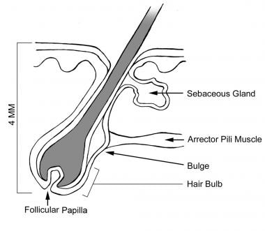
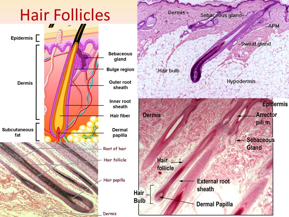
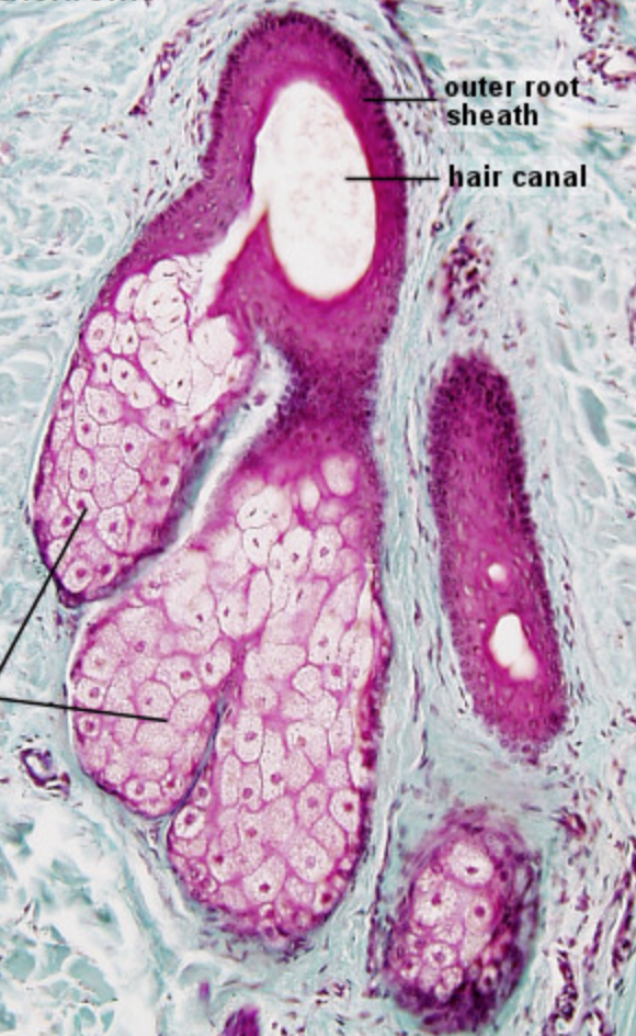
At the base of the hair folliclehair bulb there is a dermal papilla which contains the blood supply for the hair. The hair matrix which contains the proliferating cells that generate the hair and the internal root sheath is just above the dermal papilla and separated from it by a basement membrane. The hair follicle is a tubular structure with an enlarged hair bulb at its base lower left.
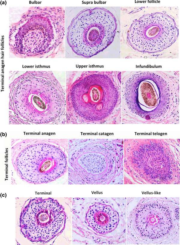
Explanation of hair and hair follicle structure and functions for hair follicle anatomy see more here. The pink speckled tissue lining the walls of the follicle is an ingrowth of the epidermis the outermost layer of the skin. During the growth phase an extra outer layer stratum basale appears.
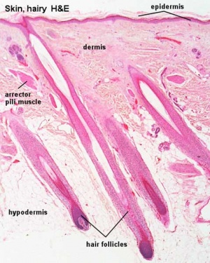
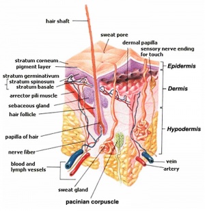
From the top down the three major skin layers shown are. Histology hair bulb view related images. Alopecia areata histology.
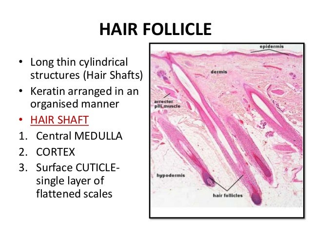
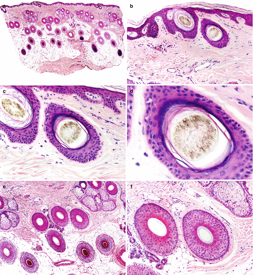
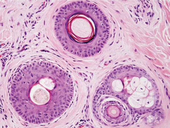
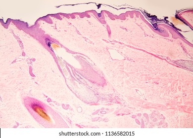
Hair histology this image shows a histological section through the adult skin showing slices through 4 hair follicles and their associated glands in different planes. Dark brown or black hair contains large ellipsoidal markedly melanized eumelanosomes whereas red hair houses spherical pheomelanosomes. The hair shaft is the yellow structure extending from the bulb within the hair follicle.
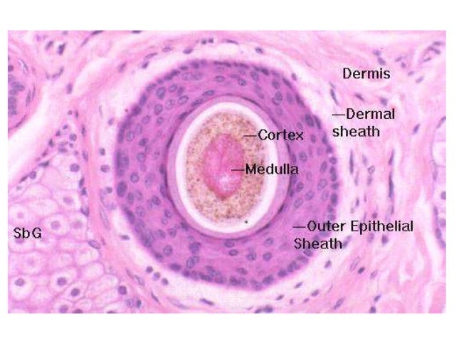
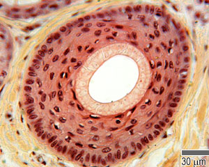
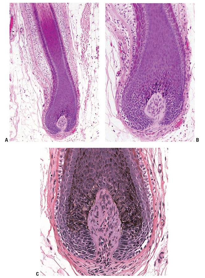
This is a section through the hair bulb at the base of a hair follicle. The hair follicles are tubular structures having a base hair bulb that surrounds the hair papilla. The hair follicle regulates hair growth via a complex interaction between hormones neuropeptides and immune cells.
Note the purple staining keratinocytes that proliferate to form the hair as well as the wall of the follicle. In gray and white hair melanocytes in bulbs are reduced in number and melanosomes are poorly melanized. The dark column of cells in the center is the hair emerging from the dead keratinocytes.
It resides in the dermal layer of the skin and is made up of 20 different cell types each with distinct functions.

 Integumentary System Hair Development Embryology
 Image Result For Skin Hair Follicle Histology Hair In 2019
 Dermal Adnexa Follicles Veterinary Histology
 Figure 1 From Integral Hair Lipid In Human Hair Follicle
 Histologic Image Of The Hair Follicle Art Print
 Dermal Adnexa Follicles Veterinary Histology
 Hair Overview 2 Low Magnification View Of An Active Hair
 Integumentary System Hair Development Embryology
 Image Result For Skin Hair Follicle Histology Hair In 2019

Presentazione Standard Di Powerpoint
 Histology Of Normal And Lpp Scalp Tissue A Structure Of
 Hair Follicle Changes Following Intense Pulsed Light
Cysts And Disorders Of The Hair
 Histology Of Human Hair Follicle Morphogenesis Sections Of

 The Hair Follicle As A Dynamic Miniorgan Sciencedirect
 Figure 2 From Controls Of Hair Follicle Cycling Semantic
 Structure And Function Of The Skin Skin Anatomy Anatomy
 Anatomy And Physiology Of Hair Intechopen
Tumours Of The Hair Follicle Dr Sampurna Roy Md
 Hair Anatomy Overview Microanatomy Of Anagen Phase Hair
 Histology Of The Skin Plastic Surgery Key
 Histologic Analysis Of Hair Follicle Growth In C57bl 6 Mice
 Anatomy And Function Of The Skin Sciencedirect
 Ahs1 Integument Hair Follicle Histology Diagram Quizlet
 Hair Follicle Changes Following Intense Pulsed Light
 Histology Of Skin Sections Top Left Epidermis Dermis
 Histology Of Wild Type And Oltnh Hair Follicles During Open I
Blue Histology Integumentary System
 110 Best Histology Skin Images Anatomy Physiology
 Skin Histology Epidermal Derivatives J F Thompson Ph D
Histology Pictorial Guide Integumentary System Pg 3
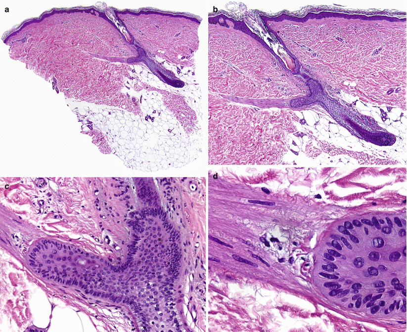 Embryology Histology And Physiology Of The Hair Follicle
:background_color(FFFFFF):format(jpeg)/images/library/3457/EZZZK6vnY0zfE0J1kUEEA_Capsule_of_Pacinian_corpuscle.png) Scalp And Hair Histology Kenhub
Scalp And Hair Histology Kenhub
 The Histological Mechanisms Of Hair Loss Intechopen
 Figure 3 From Histological Analysis Of Hair Follicle Of Dog
Blue Histology Integumentary System
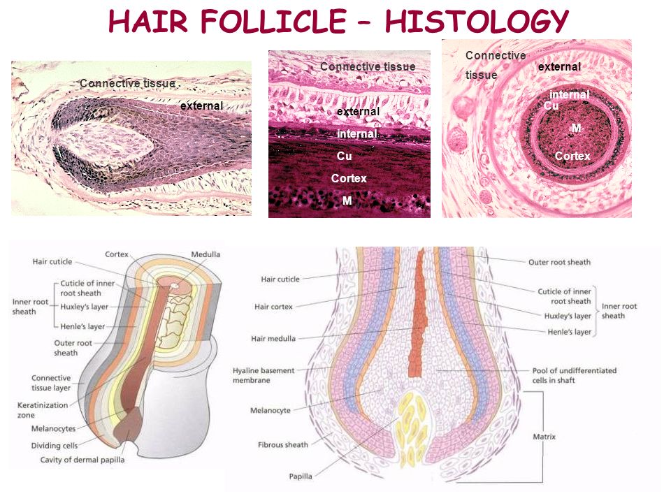 Skin Hair Nail And Mammary Gland Ppt Video Online Download
 Anagen Maturation Is Impaired In Sgk3 Null Hair Follicles
Pathology Outlines Normal Adnexae
 Hair Bulb With Papilla Hair Follicle Photos Closeup
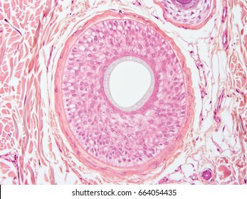 Hair Follicle Microscope Images Stock Photos Vectors
Plos One The Histological Characteristics Age Related
 Figure 1 From Histological Analysis Of Hair Follicle Of Dog
Plos Genetics Circadian Clock Genes Contribute To The
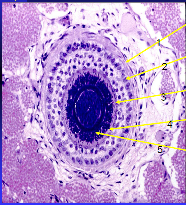
Cysts And Disorders Of The Hair
 Skin Junqueira S Basic Histology Text And Atlas 15e
The Hair Follicle Barrier To Involvement By Malignant Melanoma
:watermark(/images/watermark_only.png,0,0,0):watermark(/images/logo_url.png,-10,-10,0):format(jpeg)/images/anatomy_term/arrector-pili-muscle-3/s4X5fvj7hls141MUCJ588g_Arrector_pili_muscle.png) Scalp And Hair Histology Kenhub
Scalp And Hair Histology Kenhub
 Embryology Histology And Physiology Of The Hair Follicle
 Inflammatory Diseases Of Hair Follicles Sweat Glands And
Tumours Of The Hair Follicle Dr Sampurna Roy Md
 Histological Analyses Of A Natural Hair Follicle Isolated
Quantitative Evaluation Of Transverse Scalp Sections
Sgk3 Links Growth Factor Signaling To Maintenance Of
 Integumentary System Hair Development Embryology
Blue Histology Integumentary System
Quantitative Evaluation Of Transverse Scalp Sections
 Hair Follicle An Overview Sciencedirect Topics
:background_color(FFFFFF):format(jpeg)/images/library/3906/pSFjbYozPOhFfvjtl5qdw_Hair_follicle.png) Skin Appendages Histology Of The Nails Glands And Hair
Skin Appendages Histology Of The Nails Glands And Hair
 Sebaceous Gland Anatomy Britannica Com
 Hair Follicle Structure Anexa Beauty
 Hair Follicle Microscope Images Stock Photos Vectors

